
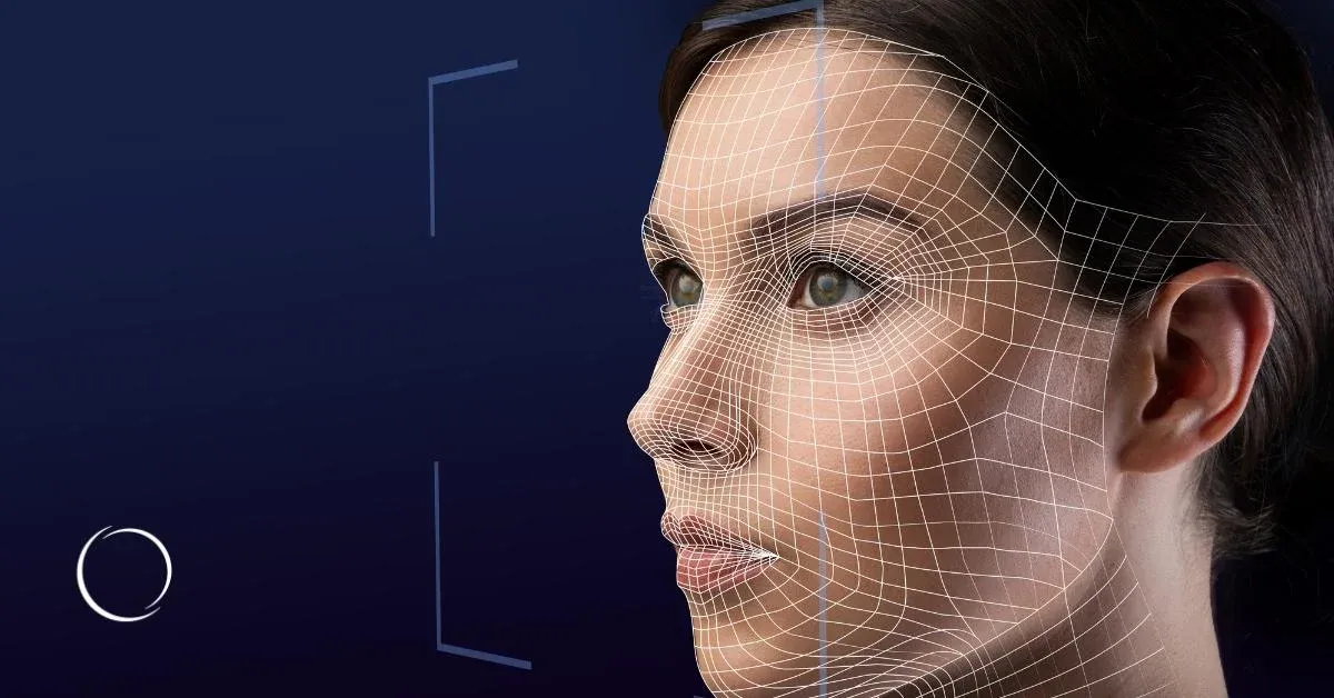
What is 3D face and body reconstruction?
3D face and body reconstruction is the process of creating a three-dimensional representation of a person's external appearance from images captured at different viewpoints. In medical and aesthetic practice, reconstructed models can support visualization, patient communication, surface measurement, and longitudinal documentation without independently diagnosing a medical condition.
From 2D photographs to interactive 3D models
Conventional photographs capture appearance from a particular viewpoint. A reconstructed 3D model represents surface geometry and texture, allowing users to inspect the model from different angles and compare compatible datasets.
Rayanix combines multi-view computer vision, camera calibration, and 3D reconstruction to develop workflows for facial and body imaging. Depending on the application and capture conditions, sparse views—such as front, left, and right images—can support textured mesh generation with real-world metric scale.
The objective is not simply to create a visually convincing image. It is to establish a repeatable capture and reconstruction workflow suited to the intended consultation, documentation, or research task.
Explore a reconstruction workflow designed around your clinical, technical, and operational requirements.
Clinical and aesthetic use cases
3D medical and aesthetic imaging provides an interactive representation of external anatomy that can support consultation, documentation, and visualization. Its appropriate role depends on the clinical question, capture protocol, reconstruction quality, and validation evidence for the intended use.
1. Plastic surgery simulation and consultation
Create patient-specific facial models for consultation and proposed-change visualization. Potential applications include rhinoplasty-related visualization, chin adjustment, jawline modification, and facial symmetry exploration. Simulations illustrate hypothetical changes; they do not guarantee surgical outcomes or predict all post-operative effects.
2. Post-operative comparison and progress tracking
Compare baseline and follow-up models using consistent capture conditions, coordinate alignment, and defined measurement methods. Meaningful comparison requires accounting for pose, facial expression, camera setup, and reconstruction variability.
3. Body contouring and volumetric measurement
Reconstruct external body surfaces for contour visualization and, where validated, dimensional or volume-related comparisons. Measurement reliability depends on the anatomical region, capture protocol, and validation of the specific metric.
4. Craniofacial and maxillofacial planning support
Use external facial surface models in visualization, research, or planning workflows. Integration with CT, CBCT, dental scans, or other clinical datasets requires separate coordinate registration and validation.
5. Dermatology and aesthetic documentation
Establish structured visual records of external facial appearance. Surface geometry and texture may complement other documentation, but a 3D model alone does not diagnose skin pathology or replace clinical examination.
6. Research, training, and education
Produce reusable 3D assets for visualization studies, research prototypes, teaching demonstrations, and medical-technology development. Downstream use depends on format compatibility, data provenance, and appropriate permissions.
Request a 3D demo or discuss your clinical workflow.
Tell us whether you need surgical visualization, longitudinal surface comparison, body scanning, or a research-oriented reconstruction pipeline.
How does the 3D reconstruction workflow work?
A multi-view 3D reconstruction workflow converts images from different viewpoints into a textured three-dimensional surface. Camera calibration, geometric reconstruction, texturing, and scale control determine the quality and usefulness of the resulting model. Each stage must be designed and evaluated for the intended visualization or measurement task.
1. Image capture
Capture facial or body images from multiple viewpoints using a suitable camera setup. A sparse-view workflow may use front, left, and right views; additional views can be considered when occlusion or surface detail requires them.
2. Camera calibration
Estimate camera parameters and relative positions. Bundle adjustment jointly optimizes camera parameters and reconstructed geometry to improve geometric consistency across views.
3. 3D geometry reconstruction
Recover surface geometry from multiple views and generate a mesh—a connected representation of the reconstructed surface. Parametric face or body models can provide structured geometry for suitable applications.
4. Texturing and metric scaling
Map image appearance onto the surface and establish real-world scale using an appropriate reference or calibrated capture setup. Texture quality and geometric accuracy must be evaluated separately.
5. Export and viewing
Deliver compatible 3D assets for downstream applications or inspect models in an in-browser viewer. Additional simulation, measurement, and clinical-system integration must be scoped separately.
Technical capabilities and specifications
Rayanix supports multi-view 3D reconstruction, metric scaling, camera calibration, parametric modeling, and standard 3D asset delivery. Quantitative performance depends on the selected hardware, capture protocol, reconstruction pipeline, and validation method; numerical specifications should be published only after engineering verification.
| Parameter | Specification |
|---|---|
| Reconstruction | Multi-view image-based 3D reconstruction |
| Capture | Sparse-view configurations, subject to application requirements |
| Geometry | Textured 3D meshes |
| Scale | Real-world metric scaling |
| Calibration | Camera calibration with bundle adjustment |
| Model refinement | Parametric modeling and differentiable-rendering-based refinement |
| Export formats | GLB, glTF, OBJ, STL, PLY |
| Visualization | Local or In-browser 3D viewing |
Benefits for clinics and patients
A well-designed 3D imaging workflow can make consultations easier to understand, provide structured records of external appearance, and enable consistent comparisons between compatible scans. These benefits depend on reliable acquisition, clear communication of simulation limitations, and an appropriate clinical workflow.
Clearer consultation: Inspect a model from multiple viewpoints rather than relying exclusively on selected photographs.
Structured documentation: Maintain standardized surface models alongside appropriately managed clinical records.
Measurable comparison: Support dimensional or surface comparisons where the method and error limits have been validated.
Adaptable workflows: Scope capture, processing, visualization, and export around operational requirements.
Reusable digital assets: Use compatible models in visualization, research, and development workflows.
These are potential workflow benefits, not guarantees of improved treatment outcomes, patient satisfaction, or diagnostic performance.
Integration and deliverables
Rayanix defines deliverables according to the clinic's existing equipment, software environment, and intended use. A project may focus on reconstruction or include calibration, model export, and interactive viewing. Integration with electronic medical records, PACS, or other clinical systems requires separate technical assessment.
Potential deliverables include:
A configured multi-view reconstruction pipeline.
Textured 3D models in GLB, glTF, OBJ, STL, or PLY.
An in-browser 3D viewer, subject to the agreed scope.
Camera calibration and capture-protocol documentation.
A technical validation report with agreed metrics, if included.
Integration interfaces or deployment components, where separately scoped.
Deployment options: On-premises processing, cloud deployment, and hybrid architecture are to be confirmed for each project. Hardware compatibility, operating environment, data transfer, maintenance, and security responsibilities must be agreed before implementation.
Regulatory and ethical positioning
The solution supports visualization, documentation, and specified planning workflows; it does not independently diagnose conditions, prescribe treatment, or replace qualified clinical judgment. Regulatory classification depends on the intended purpose, actual functionality, product claims, and jurisdiction in which the solution is marketed or used.
Before clinical deployment, assess applicable medical-device requirements, privacy laws, validation needs, and professional responsibilities in the target market. No FDA clearance, CE marking, or other regulatory authorization is claimed.
Why Rayanix?
Rayanix develops computer vision, 3D reconstruction, and AI/ML systems using an engineering-led approach. For medical and aesthetic imaging, the technical focus spans camera calibration, geometric reconstruction, model refinement, and downstream viewing. Project scope is defined around the customer's requirements rather than assumed to fit a standard off-the-shelf configuration.
3D reconstruction expertise: Multi-view reconstruction, metric scaling, textured meshes, and parametric modeling.
Calibration engineering: Camera calibration and bundle adjustment for multi-camera setups.
Application-specific delivery: Requirements-led scoping for clinical, research, and technical teams.
Regional accessibility: Consultation-led engagement for Iran and international markets, subject to project and deployment requirements.
The first step is a technical discussion to establish the intended use, required evidence, hardware constraints, and integration needs.
Request a 3D imaging consultation
Evaluate whether 3D face or body reconstruction fits your clinic, device, or research workflow. Tell Rayanix what you need to visualize, measure, document, or integrate so the technical approach can be scoped to your requirements.